
18 June 2025
What this article covers
Performing ERCP for jaundice patients confirms and treats the causes when imaging shows a blocked bile duct. Obstructive jaundice arises from a blocked bile duct, and the most commonly identified causes are bile duct stones, biliary stricture, cholangitis, or in some cases pancreatic or bile duct cancer. When imaging shows a blockage, ERCP aims to restore bile flow, alleviate the symptoms of jaundice and guide the next step in care. If you have been referred for ERCP, or if you or a family member has developed jaundice and wants to understand what the investigation and treatment pathway looks like, I hope this article helps. Jaundice is a symptom, not a diagnosis. Before any treatment can be decided, the cause must be identified and the cause determines everything. In this article, Dr. Appou explains:
Jaundice, the yellowing of the skin and the whites of the eyes, is not a diagnosis. It is a symptom and one that always needs investigating before any decision about treatment can be made. When patients come to me with jaundice, the first question I ask is not how do we treat this but where is this coming from. The answer to that question determines everything that follows.
Patients with jaundice typically fall into one of two groups, obstructive and non-obstructive jaundice. In obstructive jaundice, there is a physical blockage in the bile duct caused by a stone, a stricture, or a tumor, and this often requires procedural or surgical treatment. Non-obstructive jaundice comes from the liver itself where the bile ducts are clear. The management and treatment of each is completely different.
Non-obstructive jaundice occurs when the bile ducts are physically clear but something is interfering with how the liver processes bilirubin. Common causes include viral hepatitis, drug-induced liver injury, metabolic conditions, and autoimmune liver disease. These cases are typically managed medically by a gastroenterologist or hepatologist. No procedure on the bile ducts is needed.
Obstructive jaundice is where something is physically blocking bile flow. This is where a surgeon becomes involved. The most common causes I see in practice are:
Jaundice alone is a reason to seek medical advice. The following symptoms alongside yellowing of the eyes or skin mean that assessment should not be delayed:
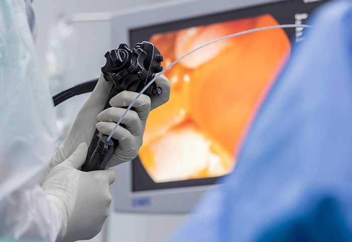
ERCP stands for Endoscopic Retrograde Cholangiopancreatography. The name is a mouthful, but the concept is straightforward: a flexible camera (endoscope) is passed through the mouth, down the esophagus and stomach, into the duodenum, the first part of the small intestine, where the bile duct and pancreatic duct open. From there, the surgeon can access the ducts directly, inject dye to make them visible on X-ray, and perform a range of treatments.
What makes ERCP particularly valuable is that it is both diagnostic and therapeutic. In many cases, the cause of the blockage is identified and treated in the same procedure, without open surgery. Depending on what is found, any of the following may be performed:
This is the most common reason to perform ERCP. A stone that has slipped out of the gallbladder and lodged in the common bile duct blocks bile flow, causes jaundice, and can trigger infection if left. ERCP is usually my first move, I pass the scope, confirm the stone, open the duct opening with a small incision, and remove the stone. All in one session, under sedation, without a surgical incision.
If stones are also present in the gallbladder, the standard approach is to clear the duct with ERCP and then remove the gallbladder laparoscopically. In many cases both steps can be completed within the same admission.
I should mention that in selected cases, where a surgeon has the appropriate advanced laparoscopic skills, bile duct exploration can be performed laparoscopically at the same time as the cholecystectomy. Using a camera passed via the cystic duct to retrieve the stones directly is an alternative to ERCP as the first step, rather than a second-line option. This is something I can discuss with patients on an individual basis.
“ERCP is usually my first move. I pass a scope, confirm the stone, open the duct opening, and remove the stone, all in one session. If the patient also has cholangitis, an infection in the bile duct, which can make people very unwell very quickly, then ERCP becomes urgent. That is not a situation to wait on.” Dr. Appou
When jaundice is caused by a tumor compressing or obstructing the bile duct, the cause is most commonly pancreatic cancer, cholangiocarcinoma (bile duct cancer), or cancers at the ampulla or duodenum, the decision about whether to place a stent via ERCP becomes more nuanced.
A stent is a small, hollow tube, think of it as a small rigid straw, placed inside the bile duct to hold the walls open and allow bile to flow freely. Plastic stents are appropriate for short-term use. Metal stents (SEMS) are generally preferred where longer-term relief of symptoms is needed. SEMS remain functional for longer before requiring replacement. All stents are inserted during ERCP under sedation.
The honest answer to ‘what treatment do I need?’ is: it depends on what is causing the blockage, whether there is infection, and whether surgery is possible. Those three questions guide every decision I make. Cancer care at this level is best delivered by a multidisciplinary team, an HPB surgeon, oncologist, radiologist, and dietitian, working together on each case.
“When the blockage is caused by a tumor, a cancer of the pancreas, the bile duct, or the surrounding structures, the decision becomes more nuanced. If the cancer is resectable, meaning we believe we can remove it completely with a view to complete surgical removal where appropriate, then I would generally want to avoid placing a stent before the operation unless the jaundice is very severe, there is active infection, or the patient needs chemotherapy first. Stenting before a resection adds a small but real risk of a complication arising. If the cancer cannot be removed, then a stent to relieve the obstruction becomes the treatment, not to cure, but to restore bile flow, protect the liver function, and relieve the itching and discomfort that obstructive jaundice causes. In that setting, a metal stent is preferable to a plastic one for most patients, because it holds the duct open for a longer period of time.”Dr. Appou
A less common but important indication is when patients who develop a bile leak as a complication of laparoscopic gallbladder removal. These patients typically present with abdominal pain and abnormal liver blood tests in the days or weeks after surgery. Comprehensive imaging, MRI or CT, usually precedes referral, to confirm the diagnosis and plan management before ERCP is performed.
Some patients ask why ERCP is recommended over other options: PTC (Percutaneous Transhepatic Cholangiography) or direct surgical exploration. The answer is about risk and recovery.
PTC involves placing a needle through the skin and into the liver under local anesthesia and sedation, guided by imaging, to access and drain the bile duct from outside. It is performed by an interventional radiologist and it carries its own complication profile, including bleeding, bile leak, worsening infection, and a risk of mortality; all factors that should be discussed with the treating team. PTC is usually reserved for cases where ERCP has failed or is not technically possible.
If ERCP cannot be completed or is unsuccessful, the options are PTC or surgical exploration. The pathway is complex, and the right choice depends on the individual case. This is a detailed discussion to have with your specialist.
ERCP is a safe procedure in experienced hands, but it is not risk-free. Complications include infection, bleeding, perforation of the duodenum or bile duct, and post-ERCP pancreatitis. There is a small, less than one percent, risk to life. These figures need to be understood in context: the alternatives to ERCP for a blocked bile duct also carry risks, often higher ones.
The complication I am asked about most often is post-ERCP pancreatitis. I want to be transparent about this, because it is also the complication that sometimes causes patients, surgeons, and gastroenterologists to hesitate about the procedure and in doing so, choose alternatives that may carry equivalent or greater risk.
“In my practice, the risk of post-ERCP pancreatitis is around 4–6%. A small group of those who develop pancreatitis may progress to severe pancreatitis, which can lead to multi-organ failure and carries a risk to life. It is very difficult to predict who will develop pancreatitis or how severe it will be. This is something I discuss openly with every patient before proceeding.”Dr. Appou
The pancreatic duct and the bile duct open close together, sometimes sharing a common channel before entering the duodenum. When the endoscope approaches the bile duct opening, there is a risk of inadvertently passing the guidewire into the pancreatic duct instead of the bile duct. If contrast dye is injected into the pancreatic duct, the risk of pancreatitis increases. Even a small amount of contrast in the pancreas raises the risk. In my practice, the steps I take to reduce this risk include:
Even with all precautions, a small residual risk remains. Every patient considering ERCP should have an open conversation with their clinician about the incidence of complications and specifically about what those rates are when the procedure is performed by an experienced operator.
A surgeon who can answer these questions directly and with confidence is one who has performed the procedure enough times to know their own outcomes.
ERCP is rarely the final step in treatment. It is one part of a management pathway. What happens next depends on what was found and what was done:
I perform ERCP at Fakeeh University Hospital, a HIMSS Stage 7-certified tertiary hospital in Dubai Silicon Oasis. FUH has a fully equipped endoscopy suite with fluoroscopy, post-procedure monitoring, a dedicated surgical team, and access to advanced imaging when needed.
I have been performing ERCP since my NHS training and have continued to develop the procedure throughout my career including attending the Scottish ERCP Masterclass, the ERCP Symposium at Salisbury Royal Infirmary, and advanced biliary courses at Colchester and Airth Castle. I have also trained other surgeons and specialty doctors in ERCP. Over 35 years of surgical practice, ERCP has become an integral part of how I manage complex biliary disease.
I consult in English and Tamil. Patients from across south-east Dubai, including DSO, Dubailand, Academic City, Mirdif, Nad Al Sheba, Mudon, and Arabian Ranches, can reach FUH directly. Appointments can be booked online via the FUH live booking link at Okadoc or contact Dr. Appou by WhatsApp or phone on +971 503 567 569 before booking.
This article is part of a wider bile duct section on drappou.com covering gallstones, bile duct cancer, choledocholithiasis, and biliary strictures. If you have a related question not covered here, I am happy to address it at consultation.
This article is written for general educational purposes and does not constitute medical advice, diagnosis, or treatment. Individual surgical decisions must be made in consultation with a qualified healthcare professional following full clinical assessment. If you are experiencing symptoms described in this article as urgent, please seek medical attention promptly. Dr. Appou Tamijmarane is licensed by the Dubai Health Authority (DHA License No: 50530660-001) and practices as a Consultant General Surgeon at Fakeeh University Hospital, Dubai Silicon Oasis, UAE. Patient testimonials referenced elsewhere on this website are published with consent in accordance with DHA advertising guidelines.
Chat